
The ABCDE rule is the international clinical benchmark used by board-certified dermatologists to detect melanoma at its most curable stage. If a pigmented lesion exhibits Asymmetry, Border irregularity, Color variegation, a Diameter over 6 millimeters, or active Evolution over time, it warrants prompt clinical evaluation. Catching atypical melanocytic proliferation before it penetrates past the epidermal basement membrane yields a 5-year survival rate exceeding 99%.
Executive Summary & Research Finding
Clinical guide to the ABCDE skin cancer rule: identify asymmetry, irregular borders, color shifts, diameter, and evolving moles before they become dangerous.
Understanding the ABCDE Criteria for Melanoma Screening
Normal benign nevi (moles) typically maintain radial symmetry, uniform brownish pigmentation, and stable dimensions across years. In contrast, cutaneous melanoma arises from malignant transformations of melanocytes, resulting in erratic microscopic growth patterns.
| Criterion | Benign Characteristics | High-Risk Malignant Indicators | Clinical Action |
|---|---|---|---|
| A - Asymmetry | Symmetrical halves (mirror image) | One half fails to match the other in shape or elevation | Photograph opposite axes |
| B - Border | Smooth, sharply demarcated round edges | Scalloped, notched, blurred, or ragged margins | Inspect under polarized light |
| C - Color | Monotone uniform tan or brown pigment | Variegation: shades of brown, black, red, white, or slate blue | Flag multi-pigment zones |
| D - Diameter | Typically under 6mm (pencil eraser size) | Lesions greater than 6mm (or rapid enlargement from tiny specks) | Measure with calibrated digital scale |
| E - Evolving | Static over decades without structural change | New itching, bleeding, crusting, elevation, or pigment spread | Urgent dermatoscope biopsy |
Step-by-Step: How to Conduct a Full-Body Skin Exam at Home
- Position yourself in a brightly lit room with a full-length mirror and a handheld hand mirror for blind spots.
- Systematically inspect head and scalp: Part hair using a blow dryer or comb, examining the neck and temples.
- Scan upper extremities: Check palms, under fingernails, between fingers, and along inner forearms.
- Inspect torso and back: Face the full-length mirror for chest and abdomen, then use the hand mirror to scan upper and lower back.
- Examine lower extremities and soles: Check backs of thighs, calves, soles of the feet, and between toes where acral lentiginous melanoma develops.
The 'Ugly Duckling' Sign (Le Vilain Petit Canard)
In addition to the ABCDE criteria, dermatologists rely on the 'Ugly Duckling' clinical concept. Most individuals produce moles that share a consistent 'signature' look. A mole that looks noticeably different, darker, or more inflamed than all your other surrounding moles should be prioritized for examination, even if it is small.
Why Manual Memory Fails: The Critical Importance of 'Evolving'
Among all five letters, E (Evolving) is clinically recognized as the single most predictive factor for malignant melanoma. The human visual cortex cannot reliably recall whether a 4-millimeter pigmented spot expanded by 0.8 millimeters over a six-month window. Without objective baseline photographs, subtle changes in borders, radial spreading, and surface texture go unnoticed until late-stage ulceration occurs.
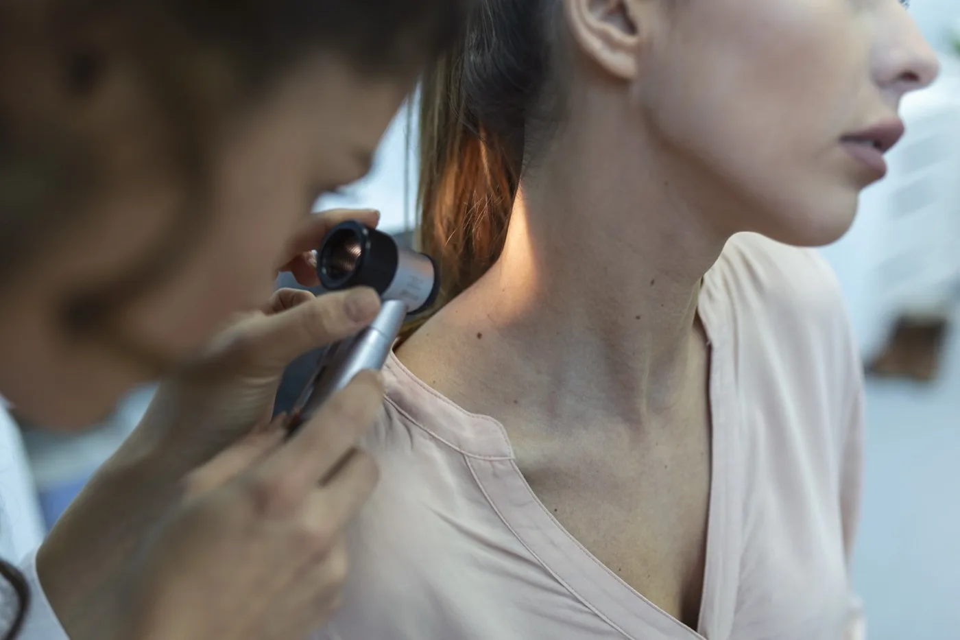
How Can Smartphone Technology Improve Home Skin Monitoring?
Modern high-resolution iPhone camera sensors combined with on-device computer vision allow users to maintain a full anatomical 3D body map. Using dedicated utilities like Mole Tracker, individuals can log macro photographs of individual lesions, document exact millimeter measurements, and receive automated calendar reminders every 90 days. Because all photos remain encrypted locally on your device, you preserve complete medical privacy while generating objective comparison logs for your dermatologist.
When Should You See a Board-Certified Dermatologist Immediately?
You should schedule an urgent evaluation if you observe spontaneous bleeding without trauma, continuous itching, rapid color darkening into jet-black or grey-blue, or satellite pigment spots appearing adjacent to an existing mole. Remember: smartphone apps are screening and tracking aids, not diagnostic instruments. If in doubt, a professional dermoscopy takes less than 15 minutes and can save your life.
Clinical ABCDE Mole Risk Assessment Checklist
Evaluate your skin lesions using the international ABCDE dermatological criteria to determine whether a spot requires professional dermatoscope evaluation.
Capture high-resolution photos with Mole Tracker to create a standardized timeline. Always consult a dermatologist for any suspicious skin changes.